neuroarchitecture of the butterfly's COMPASSTo unravel how compass information is encoded in the brain we study the monarch butterflies brain anatomically, using immunohistochemical techniques combined with imaging and 3D modelling. By staining brains with an anti-synapsin antibody, we can visualize the brain areas in the monarch butterfly brain. In addition, tracer injections into specific brain areas or into single neurons, and stainings against neurotransmitters (e.g. serotonin) allow us to gain a detailed knowledge of the neuroarchtecture of the monarch butterfly brain in general, and the compass network in particular. The brains are then imaged on a confocal laser scanning microscope and the obtained image stacks are processed in a program that allows to model brain areas or neurons in 3D. One main focus of our studies is the analysis of the central complex, the brain area that acts as the butterfly's internal complass during migration.
SPECIFIC QUESTIONS
PARTICIPATING SCIENTISTS |
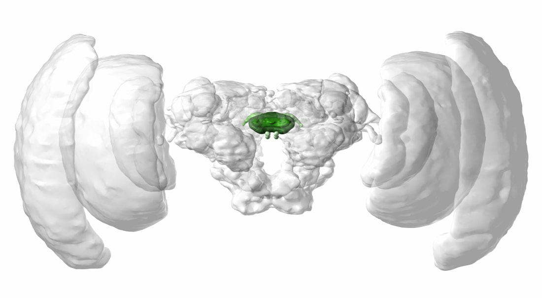
Frontal view of a 3D model of the monarch butterfly brain (the central complex is highlighted in green). From Heinze and Reppert (2012) J Comp Neurol. 3D model generated via InsectBrainDataBase.
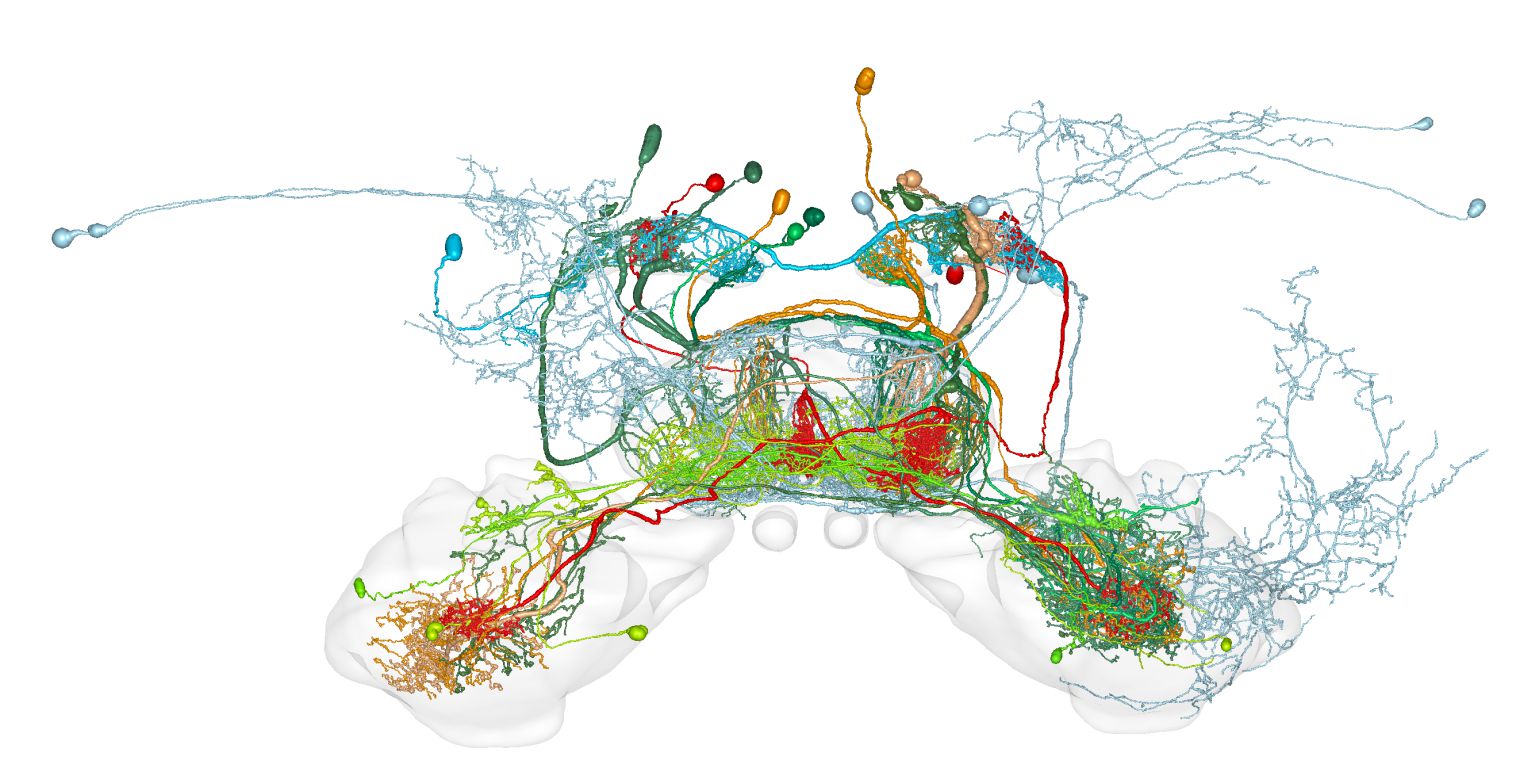
3D model of the standardized monarch butterfly central complex. Single neurons are warped into it. From Heinze et al. (2013) J Comp Neurol.
|